Débit urinaire vs. créatinine

Débit urinaire vs. créatinine :
pourquoi le facteur temps
est crucial dans la détection de la LRA

Méta-description :

La créatinine sérique est un marqueur tardif de l’atteinte rénale. Découvrez pourquoi le débit urinaire reste la mesure la plus réactive et exploitable pour la détection précoce de la LRA — en particulier en soins critiques.

Introduction

Lorsqu’il s’agit de la lésion rénale aiguë (LRA), le temps est un facteur déterminant. Plus tôt elle est détectée, plus les chances de la contrôler ou d’en inverser les effets sont grandes. Pourtant, la plupart des cliniciens s’appuient encore sur la créatinine sérique, un marqueur qui n’augmente souvent qu’une fois les lésions déjà avancées. Le débit urinaire raconte une autre histoire — et la raconte souvent en premier.

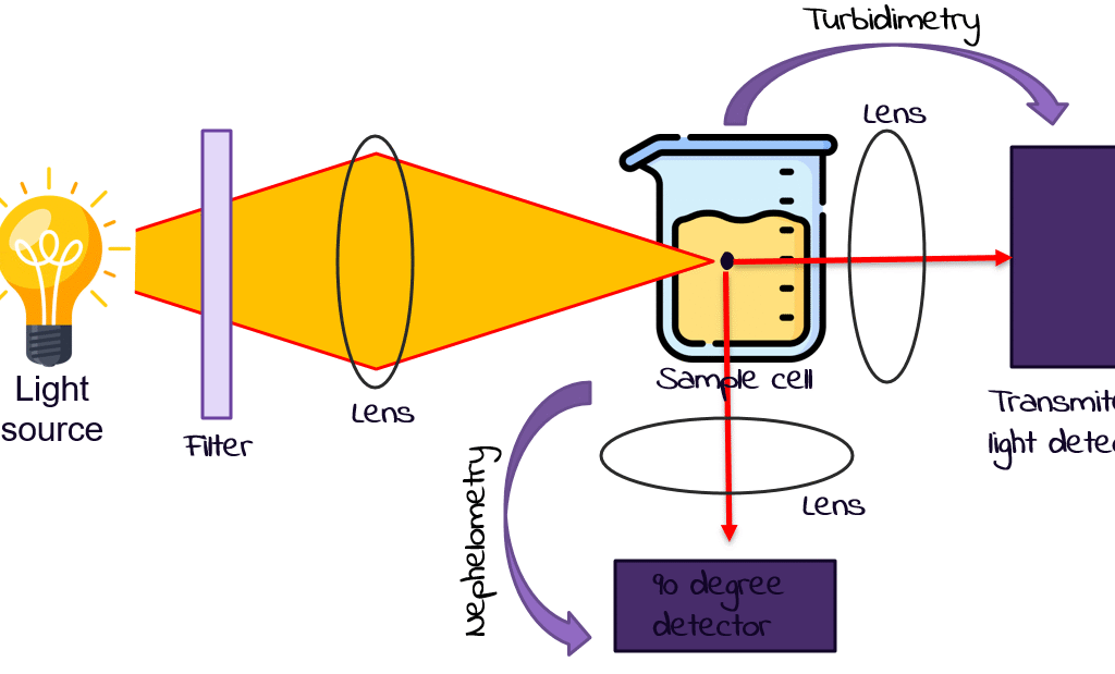
Les limites de la créatinine sérique

La créatinine sérique est facile à mesurer, mais lente à réagir. Elle peut rester dans la plage normale pendant des heures — voire des jours — après le début d’une LRA. Elle est également influencée par :
• La masse musculaire
• L’âge et le sexe
• Le niveau d’hydratation
• Certains médicaments

À l’inverse, le débit urinaire réagit en quelques heures, reflétant en temps réel la perfusion rénale et la filtration glomérulaire.

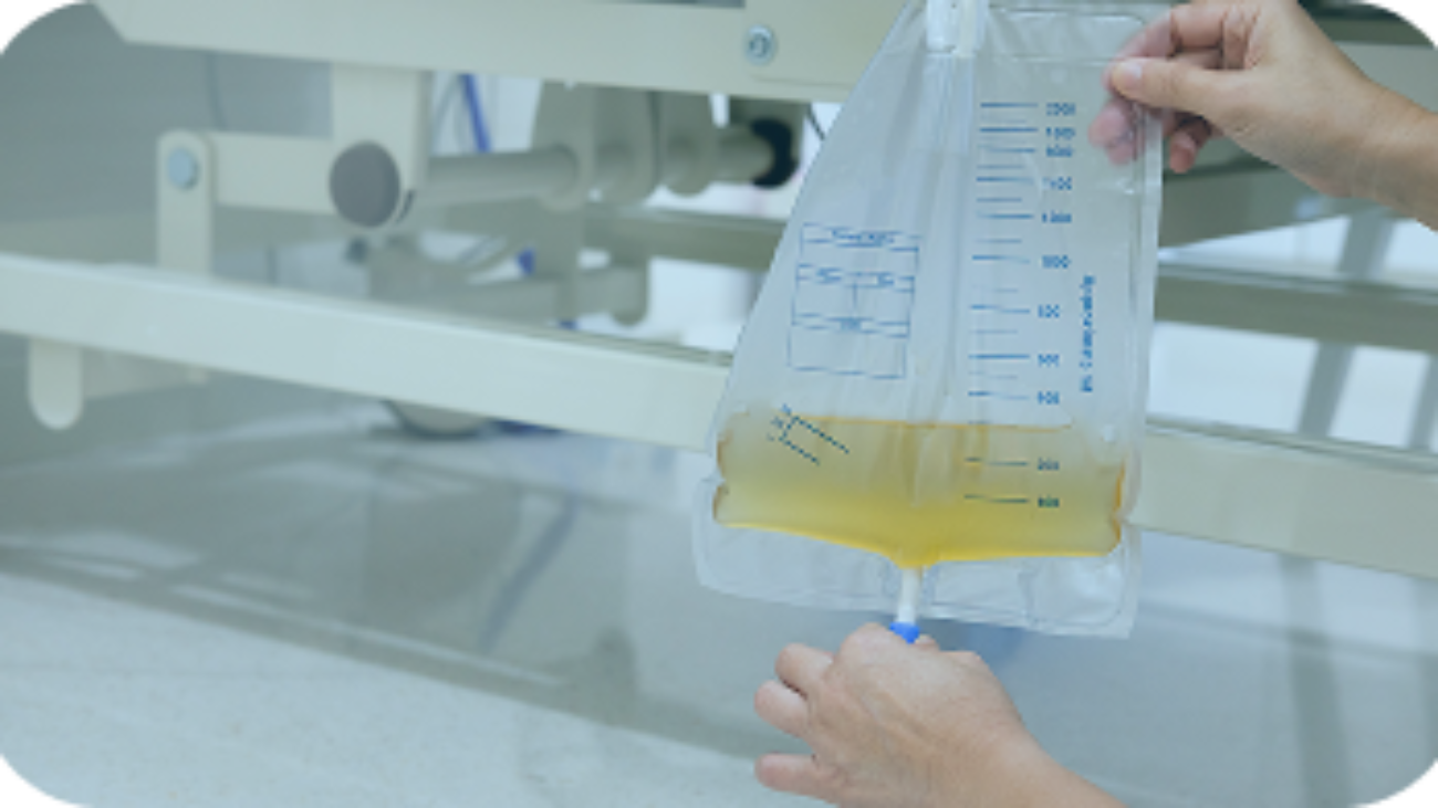
Les critères KDIGO et pourquoi le débit urinaire est essentiel

Selon les directives KDIGO sur la LRA :
• Un débit urinaire < 0,5 mL/kg/h pendant plus de 6 heures constitue un critère diagnostique de LRA — même si la créatinine est encore normale.
• De nombreuses USI passent à côté de ce signal en raison de mesures retardées, estimées ou moyennées.

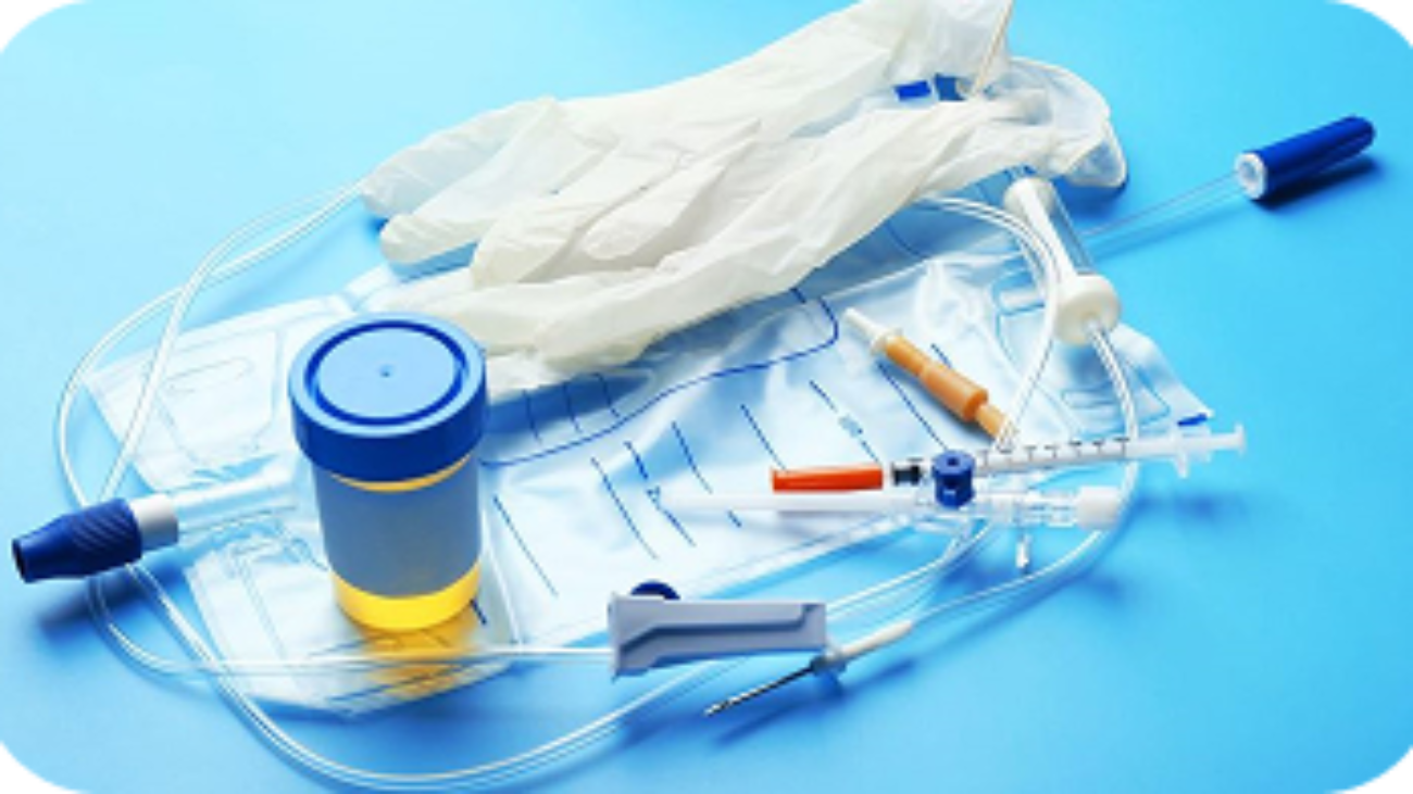
Exypnos : rendre les données de débit continues et fiables

Exypnos automatise ce paramètre critique grâce à :
• Des capteurs optiques de débit urinaire
• Un affichage en temps réel au chevet et au poste infirmier
• Des tendances historiques et des alertes précoces pour la polyurie et l’oligurie
• Une intégration avec d’autres systèmes de score clinique

Cela permet de détecter la LRA avant que des dommages irréversibles ne surviennent — et avant qu’un pic de créatinine ne confirme trop tard ce qui avait déjà été manqué.

Conclusion clinique

Pour une intervention précoce dans la LRA, le débit urinaire doit être le premier signe vital pris en compte — et non une réflexion secondaire. En soins critiques, où le retard équivaut à une détérioration, les données en temps réel sauvent des vies.

Surveillance en temps réel de la turbidité urinaire par néphélométrie

Surveillance en temps
réel de la turbidité urinairee
par néphélométrie :
un nouvel outil diagnostiqu

Méta-description :

Découvrez comment la néphélométrie permet une analyse en temps réel de la turbidité urinaire, favorisant la détection précoce des infections, des lésions rénales et le suivi de la réponse aux traitements — grâce à Exypnos.

Introduction

L’analyse d’urine est depuis longtemps un outil fondamental du diagnostic. Mais en soins critiques, la rapidité et la précision sont aussi importantes que l’exactitude. La turbidité — c’est-à-dire l’opacité de l’urine — peut révéler des infections, une hématurie et une dysfonction rénale précoce. Or, traditionnellement, elle est jugée à l’œil nu. Exypnos change la donne grâce à la néphélométrie sans contact.

Qu’est-ce que la turbidité urinaire et pourquoi est-elle importante ?

La turbidité urinaire est causée par la présence de particules telles que :
• Les globules blancs et rouges (infection ou hématurie)
• Les cristaux (calculs rénaux ou troubles métaboliques)
• Les bactéries et le mucus (infections urinaires)

Si les tests en laboratoire offrent de la précision, ils arrivent souvent trop tard pour permettre une intervention précoce. En USI, la détection en temps réel est nécessaire pour réagir avant la détérioration.

Comment fonctionne la néphélométrie dans la surveillance urinaire

La néphélométrie mesure la façon dont les particules diffusent la lumière. Dans Exypnos, un système de diffusion laser à 90° capture les valeurs NTU (unités de turbidité néphélométrique) à mesure que l’urine s’écoule dans une chambre stérile.

Les avantages de la néphélométrie incluent :
• Analyse sans contact (réduction du risque d’infection)
• Précision avec une marge d’erreur de ±2 %
• Détection immédiate des pics de turbidité (ex. apparition d’une hématurie)
• Suivi continu plutôt qu’un simple échantillonnage ponctuel

Applications cliniques réelles

La surveillance néphélométrique de la turbidité aide à :
• Détecter les infections urinaires et les infections associées aux cathéters (CAUTI)
• Suivre les lésions rénales chez les patients oncologiques ou septiques
• Valider la réponse aux traitements (ex. diurétiques)
• Dépister les populations à haut risque (LRA, sepsis, chimiothérapie)

Elle soutient également le diagnostic prédictif lorsqu’elle est associée à une logique d’IA, en identifiant des schémas bien avant que le clinicien ne voie apparaître les symptômes.

Pourquoi c’est indispensable en soins intensifs

Les soins intensifs exigent des données rapides, quantifiables et à faible risque. L’analyse de la turbidité a longtemps été trop lente, trop manuelle ou trop invasive. Exypnos la rend automatique et exploitable, en ajoutant une puissance diagnostique sans perturber les flux de travail.

Conclusion

Avec Exypnos, la turbidité n’est plus une estimation subjective. C’est un flux de données — clair, continu et cliniquement pertinent. Grâce à la néphélométrie, nous offrons aux équipes soignantes un signal supplémentaire — un signal qui pourrait bien arriver assez tôt pour changer l’issue.

Le coût clinique de la surveillance manuelle

Le coût clinique de la
surveillance manuelle
du débit urinaire en soins critiques

Méta-description :

La surveillance manuelle du débit urinaire en soins intensifs est sujette aux retards, aux erreurs et aux risques. Découvrez pourquoi des systèmes automatisés comme Exypnos sont essentiels pour la détection précoce de la LRA et une meilleure prise en charge des patients.

Introduction

Dans l’environnement à enjeux élevés des soins critiques, où chaque décision peut affecter la survie, la précision et la rapidité des données sont cruciales. Pourtant, l’un des indicateurs les plus importants de la fonction rénale — le débit urinaire — est encore suivi manuellement dans de nombreuses unités de soins intensifs (USI) à travers le monde. Cette approche dépassée introduit des risques que l’automatisation moderne peut éliminer.

Pourquoi le débit urinaire est-il important ?

Le débit urinaire est un marqueur sensible et en temps réel de la perfusion rénale, de l’équilibre hydrique et du fonctionnement des organes. Il joue un rôle clé dans :
• La détection précoce de la lésion rénale aiguë (LRA)
• L’évaluation des effets des diurétiques et des vasopresseurs
• L’orientation de la réanimation liquidienne chez les patients septiques ou postopératoires

Selon les critères KDIGO, une baisse du débit urinaire (oligurie) précède souvent les changements de créatinine sérique chez les patients atteints de LRA. Cela fait de la surveillance continue du débit urinaire non seulement une aide, mais une nécessité.

Le problème du suivi manuel

Dans les flux de travail traditionnels en soins intensifs, les infirmières vérifient manuellement les poches urinaires et consignent les volumes toutes les heures. Cependant :
• Les études montrent que jusqu’à 39 % des mesures sont manquées ou retardées
• Des erreurs de 17 ml/heure sont courantes — suffisantes pour franchir les seuils diagnostiques de la LRA
• Les méthodes manuelles augmentent le risque de contamination et la charge de travail

Ces limites entraînent des alertes manquées, des interventions retardées et parfois des complications mettant en jeu le pronostic vital.

L’automatisation comme solution : en temps réel, fiable et peu intrusive

Les systèmes modernes comme Exypnos utilisent des capteurs optiques et des chambres stériles jetables pour automatiser l’ensemble du processus. Leurs caractéristiques incluent :
• Une analyse continue du volume urinaire et de la turbidité
• Des alertes basées sur l’IA pour les déséquilibres hydriques et les anomalies
• Une intégration fluide avec les moniteurs au chevet et les tableaux de bord du poste infirmier
• Une réduction de la charge de travail infirmière et moins d’interventions manuelles

Avec Exypnos, les données sur le débit urinaire deviennent partie intégrante d’un modèle de soins réactif, et non plus une trace papier différée.

Conclusion

Dans un domaine où le temps est un facteur vital, s’appuyer sur des estimations manuelles pour un paramètre critique comme le débit urinaire n’est plus acceptable. La surveillance automatisée, telle que celle fournie par Exypnos, favorise un diagnostic plus précoce, une meilleure gestion des fluides et, en fin de compte, des résultats plus sûrs pour les patients à haut risque.

image_2025-10-08_09-37-13 36

Exypnos Medical Joins the Hamilton–Niagara Bootcamp 2025

Exypnos Medical Joins the Hamilton
Niagara Bootcamp 2025

Toronto, Canada – October 2025

Exypnos Medical is pleased to announce its participation in the Hamilton–Niagara Bootcamp 2025, a premier international acceleration program designed to support innovative technology companies expanding into the Canadian and North American markets.

Organized by Global Startups, in partnership with the Hamilton and Niagara Economic Development Offices, the program brings together a select group of global ventures for a week of business immersion, networking, and collaboration across Ontario’s innovation corridor. The event runs from October 14 to 21, 2025, and includes participation in the Toronto Global Forum, as well as dedicated sessions in Niagara and Hamilton focused on ecosystem engagement and sector partnerships.

Participation in this program marks an important milestone in Exypnos Medical’s international growth strategy. Through this opportunity, the company aims to strengthen its connections with Canadian healthcare institutions, research centers, and manufacturing partners, paving the way for future clinical collaborations and regulatory advancement in North America.

Exypnos welcomes all participants and ecosystem partners attending the Toronto Global Forum and the Hamilton–Niagara Bootcamp to connect and exchange ideas on advancing patient monitoring technologies.

13 urin 1

Clinical and Economic Impact of CAUTIs and the Role of Contactless Monitoring

Clinical and Economic Impact
of CAUTIs and the Role of
Contactless Monitoring.

Meta Description:

atheter-associated urinary tract infections (CAUTIs) are among the most common healthcare-associated infections and are…

Introduction

Catheter-associated urinary tract infections (CAUTIs) are among the most common healthcare-associated infections and are a particular concern in intensive care units (ICUs). A CAUTI occurs when bacteria enter the urinary tract via an indwelling catheter and cause infection. Because many ICU patients require indwelling urinary catheters for hourly urine output monitoring or other reasons, CAUTIs occur disproportionately in high-acuity settings. They not only cause patient discomfort and morbidity but can lead to serious complications like urosepsis and prolonged hospital stays. This article examines the clinical and economic impact of CAUTIs in ICUs, discusses how current practices (such as manual urine output monitoring) contribute to the problem, and explores how contactless, automated urine monitoring systems (such as Exypnos) offer a safer, more reliable alternative.

CAUTI Prevalence and Clinical Burden in ICUs

Urinary tract infections are a leading type of hospital-acquired infection, accounting for an estimated 62,700 cases in U.S. acute care hospitals in 2015. Importantly, virtually all healthcare-associated UTIs are linked to urinary catheters. In ICUs the problem is amplified: at any time, 60–90% of ICU patients may have a catheter in place (compared to 10–30% on general wards), and nearly 95% of UTIs in ICU settings are catheter-associated. Infection surveillance data show ICU CAUTI incidence rates on the order of 3–7 infections per 1,000 catheter-days – several-fold higher than in lower-acuity units. Each day of catheterization poses a compounding risk (roughly a 3–7% increase in infection risk per day of indwelling time), so extended catheter use greatly elevates the chance of infection.

The clinical burden of CAUTIs in critically ill patients is substantial. A bladder infection in a fragile ICU patient can quickly escalate: CAUTIs frequently lead to complications such as pyelonephritis or gram-negative bacteremia. Indeed, CAUTIs are a leading cause of secondary sepsis in the hospital – roughly 17% of hospital-acquired bacteremias are attributed to a urinary source. If a urinary infection progresses to bloodstream infection in an ICU patient, mortality rates are high (on the order of 10–30% in reported series). Even when not causing overt sepsis, CAUTIs prolong ICU and hospital length of stay and necessitate invasive procedures (e.g. central lines for antibiotics) that can trigger further complications. The U.S. CDC estimated that over 13,000 deaths each year are associated with UTIs, mostly catheter-related. In short, CAUTIs add significant morbidity and mortality to already high-risk ICU patients, making prevention a top priority.

Economic Impact of CAUTIs

In addition to clinical harm, CAUTIs carry a heavy economic impact. Treating a single catheter-associated UTI involves extra laboratory tests, antimicrobial therapy, and often days of additional in-hospital care. Estimated costs per CAUTI case vary, but studies peg the average inpatient cost in the range of $1,000 up to $13,000 or more. Notably, ICU-acquired CAUTIs tend to be the most expensive, since these patients are often sicker and the infections more severe (one analysis found an average cost of about $10,000 for a CAUTI in ICU patients). Across the United States, the aggregate cost burden is hundreds of millions of dollars annually. These infections also incur indirect costs: they can extend ICU occupancy (blocking new admissions), require costly isolation or enhanced nursing care, and contribute to antimicrobial resistance through antibiotic use.

Because CAUTIs are considered largely preventable, payers and regulators have taken note. In the U.S., the Centers for Medicare & Medicaid Services (CMS) identifies CAUTI as a preventable hospital-acquired condition and may deny hospitals reimbursement for its treatment. This creates financial incentives for hospitals to reduce CAUTI rates. Additionally, an avoidable infection adversely affects quality metrics and can lead to financial penalties under value-based purchasing programs. In summary, every CAUTI in the ICU not only endangers the patient but also carries a price tag – one that hospitals increasingly must absorb, providing strong motivation to invest in better prevention and monitoring strategies.

Why Manual Urine Output Monitoring Contributes to Risk

One less-appreciated factor in CAUTI risk is the traditional method of monitoring urine output in ICUs. Accurate urine output measurement is vital for managing critically ill patients (for example, tracking hourly output is key for guiding fluid resuscitation and detecting renal impairment). This need for precision is a major reason so many ICU patients are catheterized. However, manual urine output monitoring introduces infection risks and can delay recognition of patient deterioration.

From an infection control standpoint, each interaction with the catheter or drainage bag is a potential entry point for bacteria. Best practices dictate maintaining a closed sterile drainage system continuously, because opening or disconnecting the system even briefly allows pathogens to enter. Studies have shown that if a urinary drainage system is left open to air (no closed circuit), the infection rate approaches 100%, whereas using a continuously closed system can keep infection rates under 25%. Consequently, frequently breaking the catheter system to empty urine or measure output will increase CAUTI risk. In many ICUs, nurses empty urometer chambers or bags hourly to record output, and each of those manipulations – even done with gloves and aseptic technique – is an opportunity for contamination.

Equally important, manual monitoring is prone to lapses in timeliness and accuracy, which can result in missed warning signs like oliguria. ICU nurses typically document urine output hourly, but in practice these readings often run behind or get batched if the unit is busy. In a study comparing electronic monitoring to nurse charting, manual urine outputs were recorded with an average delay of 47 minutes (median ~18 min), and occasionally up to 6 hours late. Nurses also tended to overestimate volumes by around 20 mL per hour compared to the actual output captured by an automated device. These delays and inaccuracies mean a drop in urine output might go unnoticed for hours. A patient could develop oliguria (low output) at 2 PM, but if staff are tied up with an emergency, it may not be measured and recognized until much later. This matters because prolonged unrecognized oliguria is dangerous – one analysis found that a delay of more than 6 hours in addressing oliguria was associated with a ~30% increase in ICU mortality risk. In summary, traditional urine monitoring forces a trade-off: checking output frequently is clinically important but doing so manually increases infection exposure and is limited by human timing. This is where automation offers a compelling solution.

Contactless, Automated Urine Monitoring: A Safer Alternative

New “contactless” automated urine monitors have been developed to mitigate the pitfalls of manual monitoring. These systems allow continuous urine output measurement without requiring hourly human intervention or any opening of the drainage system. For example, the Exypnos urine monitoring system uses an optical sensor and a closed, inline collection chamber to automatically measure urine volume in real time. The disposable chamber connects seamlessly to standard Foley catheter tubing and remains sealed, so urine flows through a sterile pathway that the device can measure without ever exposing the urine to the environment. Urine volume data are transmitted continuously to a bedside monitor or central station, giving up-to-the-minute insight into output.

Because the sensor does not require breaking the circuit or touching the urine, the closed sterile drainage is maintained at all times, dramatically reducing contamination risk. In essence, automated monitors uphold the “closed system” ideal far better than any manual method. There is no need for a nurse to open a drain port every hour – the device does the monitoring automatically. Additionally, these systems are highly accurate. In the ICU study mentioned earlier, the electronic monitor’s measurements were virtually identical to true output (within a couple milliliters), whereas nurse measurements showed significant error. This means clinicians can trust the readings and not worry about charting delays or misestimation. If a patient’s urine output drops suddenly, the change is evident immediately on the monitor, and alarms can be set to alert staff promptly.

Clinical Advantages Beyond Infection Control

Implementing contactless automated urine monitoring in ICUs provides benefits that extend beyond reducing CAUTIs. One major advantage is earlier detection of acute kidney injury (AKI) and other deterioration. Urine output is a sensitive indicator of renal perfusion – often declining hours or days before serum creatinine levels rise. Studies show that relying on creatinine alone can miss a large portion of AKI in critical care; in one analysis, serum creatinine criteria failed to identify over one-third of AKI cases that were detectable by oliguria. Moreover, even brief episodes of oliguria are prognostically significant: ICU patients who meet AKI criteria by urine output (oliguria) have been shown to have higher mortality than those who meet it by creatinine rise alone. By delivering continuous urine output trends, automated monitors enable the care team to recognize AKI in its earliest phase and intervene sooner (for example, by optimizing volume status, avoiding nephrotoxic drugs, or addressing causes of low output). This proactive approach could improve renal outcomes and overall patient survival.

Another benefit is enhanced fluid and hemodynamic management. Minute-to-minute urine output is essentially a real-time gauge of kidney perfusion and volume status. With continuous monitoring, clinicians can respond faster to a patient’s fluid needs – for instance, detecting an insidious drop in output that might signal the onset of shock or, conversely, noticing a brisk diuresis that could lead to dehydration. Research indicates that patients who undergo more intensive urine output monitoring tend to have less fluid overload and better outcomes than those monitored less frequently. Automated systems make such intensive monitoring feasible around the clock without increasing staff workload.

Finally, there are workflow and safety advantages for nursing staff. Automation frees ICU nurses from the repetitive task of measuring and emptying urine bags every hour. This not only saves time (allowing nurses to focus on direct patient care) but also reduces staff exposure to potentially infectious urine. Early implementations have shown that automated urine monitoring can streamline nursing workflow and reduce time spent on documentation and waste handling. In high-acuity environments, where nurse-to-patient ratios are tight, this efficiency gain is very valuable. Additionally, the continuous data logs can be integrated into electronic medical records, improving chart accuracy and enabling advanced clinical decision support (such as alerting if output falls below a set threshold).

Conclusion

CAUTIs impose a serious clinical and financial burden in ICUs, but innovations in urine output monitoring are helping turn the tide. By eliminating unnecessary breaks in the catheter closed system and by providing immediate awareness of changes in renal output, contactless automated monitoring systems directly tackle two key contributors to CAUTIs: contamination during manual measurements and delayed recognition of patient decline. Early studies and real-world use indicate that these technologies can reduce infection risk, enhance early AKI detection, optimize fluid management, and ease the workload on ICU staff – all without compromising the quality of patient data. For intensivists and infection control teams, such systems represent a promising strategy to improve ICU care. In an era where hospitals are increasingly accountable for preventing device-related infections, automated urine monitoring offers a compelling, evidence-based tool to improve patient safety and outcomes in high-acuity environments.

Sources:

  1. Centers for Disease Control and Prevention (CDC). Clinical Safety: Preventing Catheter-associated Urinary Tract Infections (CAUTIs). June 27, 2025.
  2. NHSN Patient Safety Component Manual: Urinary Tract Infection Events. January 2025
  3. California Department of Public Health (CDPH). CAUTI Prevention Guidelines. 2017
  4. Nicastri E, Leone S. Healthcare Associated Urinary Tract Infections – ISID Guide to Infection Control. June 2021
  5. CDC/HICPAC. Guideline for Prevention of CAUTI. 2009
  6. Cho HJ, et al. Impact of Infection Prevention Programs on CAUTI in ICUs. J Korean Med Sci. 2024
  7. NHSN Data: “UTIs are 5th most common HAI” (2015 data)
  8. E, et al. Health Care–Associated Infections: A Meta-analysis of Costs. JAMA Intern Med. 2013
  9. Additional Hospital Inpatient Cost of CAUTI – Meta-analysis. 2017
  10. ISID Guide (Nicastri & Leone). CAUTI costs and outcomes.
  11. Meddings J, et al. Effect of Nonpayment for Hospital CAUTIs. Ann Intern Med. 2012
  12. Smith A, Kellum JA, et al. Automated vs Manual Urine Output Monitoring in ICU. Crit Care. 2021
  13. Smith A, Kellum JA, et al. Urine Output and AKI Detection in ICU. Crit Care. 2021
  14. Jin K, et al. Intensive UO Monitoring and Outcomes. (Referenced in Smith et al. 2021)
  15. Exypnos Medical. Product Overview: Exypnos UO Monitoring System. 2025
  16. Exypnos Medical. Clinical Impact of Manual vs Automated Monitoring. 2025
  17. Exypnos Business Plan (user-provided data). Nursing Workflow Benefits. 2025
13

Urine Output vs. Creatinine: Why Timing Matters in AKI Detection

Urine Output vs.
Creatinine: Why Timing
Matters in AKI Detection

Meta Description:

Serum creatinine is a late marker of kidney injury. Learn why urine output remains the most responsive and actionable measure for early AKI detection—especially in critical care.

Introduction

When it comes to acute kidney injury (AKI), timing is everything. The sooner you detect it, the better your chances of reversing or controlling damage. But most clinicians still rely on serum creatinine, a marker that often rises after injury has progressed. Urine output tells a different story—and often tells it first.

The Limits of Serum Creatinine

Serum creatinine is easy to measure but slow to respond. It can remain within normal range for hours—or even days—after AKI onset. It’s also affected by:
• Muscle mass
• Age and sex
• Hydration levels
• Certain medications
In contrast, urine output responds within hours, reflecting real-time renal perfusion and glomerular filtration.

KDIGO Criteria and Why Urine Output Is Crucial

According to the KDIGO AKI guidelines:
• A urine output of <0.5 mL/kg/h for 6+ hours is an AKI diagnostic criterion—even if creatinine is still normal.
• Many ICUs miss this signal due to delayed, estimated, or averaged measurements.

Exypnos: Making Output Data Continuous and Reliable

Exypnos automates this critical parameter using:
• Optical urine flow sensors
• Real-time display at the bedside and nurse station
• Historical trends and early warning alerts for polyuria and oliguria
• Integration with other clinical scoring systems
This ensures AKI can be caught before irreversible damage occurs, and before a creatinine spike confirms what was already missed.

The Clinical Bottom Line

For early intervention in AKI, urine output should be the first vital sign considered—not an afterthought. In critical care, where delay equals deterioration, real-time data saves lives.

Picture1 11

Real-Time Urine Turbidity Monitoring with Nephelometry: A New Diagnostic Tool

Real-Time Urine Turbidity
Monitoring with
Nephelometry: A New
Diagnostic Tool

Meta Description:

Explore how nephelometry enables real-time urine turbidity analysis, supporting earlier detection of infections, kidney damage, and treatment response tracking—powered by Exypnos.

Introduction

Urinalysis has long been a fundamental tool in diagnostics. But in critical care, speed and precision are just as important as accuracy. Turbidity—the cloudiness of urine—can reveal infections, hematuria, and early kidney dysfunction. But traditionally, it’s been judged by sight. Exypnos is changing that with non-contact nephelometry.

What Is Urine Turbidity and Why It Matters

Urine turbidity is caused by particles such as:
• White and red blood cells (infection or hematuria)
• Crystals (kidney stones or metabolic disorders)
• Bacteria and mucus (UTIs)
While lab-based testing offers accuracy, it often comes too late for early intervention. In ICUs, real-time detection is needed to respond before deterioration sets in.

How Nephelometry Works in Urine Monitoring

Nephelometry measures how particles scatter light. In Exypnos, a 90° laser scattering system captures NTU (Nephelometric Turbidity Units) values as urine flows through a sterile chamber.

Benefits of nephelometry include:

• Contact-free analysis (reduces infection risk)
• Precision at ±2% error rate
• Immediate detection of turbidity spikes (e.g., onset of hematuria)
• Continuous trending, not just one-time sampling

Real Clinical Applications

Nephelometric turbidity monitoring helps in:
• Detecting UTIs and catheter-associated infections (CAUTIs)
• Monitoring kidney injury in oncology or septic patients
• Validating medication response (e.g., post-diuretic flush)
• Screening high-risk populations (AKI, sepsis, chemotherapy)
It also supports predictive diagnostics when paired with AI logic, identifying patterns long before a clinician might see symptoms.

Why It Belongs in the ICU

ICUs demand fast, quantifiable, and low-risk data. Turbidity analysis has traditionally been too slow, too manual, or too invasive. Exypnos makes it automatic and actionable, adding diagnostic power without workflow disruption.

Conclusion

With Exypnos, turbidity is no longer a subjective guess. It’s a data stream—clean, continuous, and clinically meaningful. By using nephelometry, we give healthcare teams another signal to work with—one that might just arrive early enough to change the outcome.

12 urin 1

The Clinical Cost of Manual Urine Output Monitoring in Critical Care

The Clinical Cost of
Manual Urine Output
Monitoring in Critical Care

Meta Description:

Manual urine output monitoring in ICUs is prone to delays, errors, and risks. Learn why automated systems like Exypnos are essential for early AKI detection and better patient care.

Introduction

In the high-stakes environment of critical care, where every decision can affect survival, the accuracy and timing of data are crucial. Yet one of the most important indicators of kidney function—urine output—is still tracked manually in many ICUs worldwide. This outdated approach introduces risks that modern automation can eliminate.

Why Urine Output Matters

Urine output is a sensitive, real-time marker of renal perfusion, fluid balance, and organ function. It plays a key role in:
• Early detection of Acute Kidney Injury (AKI)
• Assessing the effects of diuretics and vasopressors
• Guiding fluid resuscitation in septic or post-op patients
According to KDIGO criteria, a drop in urine output (oliguria) often precedes serum creatinine changes in AKI patients. This makes continuous urine monitoring not just helpful—but essential.

The Problem with Manual Tracking

In traditional ICU workflows, nurses manually check urine bags and record output on an hourly basis. However:
• Studies show up to 39% of measurements are missed or delayed
• Errors of 17 ml/hour are common—enough to cross AKI diagnostic thresholds
• Manual methods increase contamination risk and workload
These shortcomings lead to missed early warnings, delayed interventions, and even life-threatening complications.

The Case for Automation: Real-Time, Reliable, Low-Touch

Modern systems like Exypnos use optical sensors and sterile disposable chambers to automate the entire process. Features include:
Continuous urine volume and turbidity analysis
AI-based alerts for fluid imbalances and abnormal patterns
Seamless integration with bedside monitors and nurse station dashboards
Reduced nurse workload and fewer manual interventions
With Exypnos, urine output data becomes part of a responsive care model, not a delayed paper trail.

Conclusion

In a field where timing is everything, relying on manual estimation for a critical parameter like urine output is no longer acceptable. Automated monitoring, like that provided by Exypnos, supports earlier diagnosis, better fluid management, and ultimately, safer outcomes for high-risk patients.